As introduced in the previous blog post in this series, HLA molecules are central to the presentation of tumor antigens and anti-tumor immune recognition. Building on our previous discussion of classical and non-classical HLA molecules in healthy tissues, we now explore how tumors manipulate HLA pathways to evade immune detection and destruction. These adaptations range from loss of HLA gene expression to active exploitation of HLA-mediated immune regulatory mechanisms within the tumor microenvironment. In this blog post, we dive deeper into some of the major ways cancers leverage HLA biology to stay one step ahead of the immune system.
Expression modulation of HLA Class I and associated genes in cancer cells
Downregulation of HLA Class I expression is one of the most common mechanisms of tumor immune escape and has direct clinical consequences. When cancer cells reduce surface expression of HLA Class I proteins, via gene deletion, loss-of-function mutations, defects in antigen processing, or epigenetic silencing, they become less visible to cytotoxic CD8+ T cells. This may help explain why some patients fail to respond to, or relapse after, immunotherapies such as PD-1/PD-L1 checkpoint inhibitors [1].
Paradoxically, this same HLA Class I expression loss can increase susceptibility to natural killer (NK) cell-mediated killing. NK cells express killer cell immunoglobulin-like receptors (KIRs), a diverse family of activating and inhibitory receptors whose interaction with functional HLA molecules ensures that NK cells do not attack healthy cells (“self-tolerance”). When cancer cells downregulate HLA Class I expression, this inhibitory signal can be lost, promoting NK cell activation. This process is called “missing-self recognition”. This biology has inspired the development of NK-cell-based therapies, including CAR-NK approaches and NK-cell engagers designed to exploit HLA Class I-low tumors [2]. Numerous NK-cell-based therapies are currently being evaluated clinically, including non-engineered NK cells, CAR-NK therapies, and combination approaches [3].
Beyond HLA Class I expression loss, tumor cells exposed to DNA damage, hypoxia, or oncogenic signaling can upregulate stress-induced ligands, including MICA and MICB. These ligands activate the NKG2D receptor on NK cells and cytotoxic T cell subsets, providing a parallel immune-surveillance pathway. However, tumors can also actively downregulate or shed these ligands as an escape mechanism [4]. Consequently, therapeutic strategies aimed at restoring or enhancing MICA/B-mediated recognition, including monoclonal antibodies [5] and engineered cell therapies [6, 7], are currently under clinical investigation.
Together, these observations illustrate how alterations in HLA Class I expression can simultaneously facilitate escape from T-cell immunity and increase vulnerability to NK-cell-mediated recognition, creating both challenges and opportunities for cancer immunotherapy.
Non-classical HLA-mediated regulation in the tumor microenvironment
Beyond expression down- and upregulation of HLA Class I and HLA-associated genes, tumors employ an additional layer of immune evasion: actively recruiting and reprogramming immune cells present in the tumor microenvironment to suppress immune responses rather than attack the tumor cells. Several non-classical HLA molecules are key mediators of this immunosuppressive crosstalk.
In a striking convergence of immune tolerance and tumor immune escape, HLA-E is overexpressed in several cancer types, including colorectal, gastric, and lung cancer. HLA-E protein binds to inhibitory receptor NKG2A, which is expressed on NK cells and tumor-infiltrating CD8+ T cells. As such, HLA-E overexpression can function as an immune checkpoint, dampening anti-tumor cytotoxicity (see Figure below). Consequently, the HLA-E/NKG2A axis has emerged as an attractive therapeutic target. Monalizumab, an antibody targeting NKG2A and designed to block HLA-E-mediated inhibitory signaling, is currently being evaluated in Phase 3 trials. [8].
Similarly, HLA-G is aberrantly expressed across multiple cancer types. Through engagement of inhibitory receptors, including LILRB1 and LILRB2, and in some contexts KIR2DL4, HLA-G suppresses NK cell cytotoxicity and impairs anti-tumor immune responses. This mechanism has been implicated in resistance to trastuzumab, an antibody targeting HER2, in HER2-positive breast cancer. HLA-G is increasingly regarded as a novel immune checkpoint and a candidate therapeutic target [9].
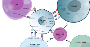
How HLA molecules interact with immune cells in the tumor microenvironment
The role of regulatory immune agents in the tumor microenvironment
Beyond directly suppressing cytotoxic lymphocytes, HLA-mediated immune evasion also extends to the myeloid compartment. HLA-G protein on the surface of cancer cells can engage the inhibitory receptors LILRB1 and LILRB2 on macrophages, promoting their differentiation into immunosuppressive tumor-associated macrophages (TAMs). The resulting TAMs produce anti-inflammatory cytokines such as IL-10 and TGF-β, suppress T-cell activation, and support tumor progression, thereby establishing a self-reinforcing immune-evasive niche. HLA Class I proteins on the surface of cancer cells also bind to LILRB1 on TAMs, inhibiting macrophage activation and phagocytic function. This HLA Class I–LILRB1 axis is now being explored as a therapeutic target, with LILRB1-blocking antibodies in pre-clinical and early clinical development. [10]. In marked contrast, HLA class II expression has been associated with reduced TAM tumor infiltration and improved clinical outcome in oropharyngeal cancer, suggesting that HLA class II-mediated immune activation may counteract myeloid-driven immune suppression [11]. Given their role in promoting tumor growth, angiogenesis, immunosuppression, and metastasis, TAMs represent an attractive therapeutic target.
Regulatory T cells (Tregs), a specialized T helper CD4+ cell subset, are a further critical immunosuppressive component of the tumor microenvironment with direct clinical relevance. Tregs accumulate in the tumor environment, where they suppress the immune activity of cytotoxic and helper T cells through inhibitory cytokines like IL-10 and TGF-β, blockade of co-stimulatory signals, and contact-dependent killing. Elevated intratumoral Treg density has repeatedly been associated with poor prognosis, including reduced overall survival, across multiple cancer types [12]. Several studies have linked tumor-associated HLA-G expression to increased Treg infiltration and adverse clinical outcomes [13]. In addition, high Treg infiltration has been associated with resistance to immune checkpoint inhibitors, making Treg quantification a potentially valuable clinical biomarker. Selective Treg-depletion strategies that target intratumoral Tregs without disrupting peripheral regulatory homeostasis are under active investigation [14]. The ability of tumors to exploit HLA-mediated pathways to recruit Tregs illustrates how immune tolerance mechanisms are co-opted to support tumor progression.
HLA genes are more than biomarkers of antigen recognition and immune activation
Taken together, these examples illustrate that HLA biology extends far beyond antigen presentation. Through modulation of classical and non-classical HLA expression, tumors can simultaneously evade cytotoxic immune recognition, suppress NK cell activity, reshape myeloid populations, and promote regulatory T cell responses. As our understanding of HLA biology continues to expand, HLA genes are increasingly emerging as more than biomarkers of immune recognition. From predicting immunotherapy response to identifying new therapeutic vulnerabilities, HLA-related pathways are becoming central to the next generation of precision immuno-oncology.
Stay tuned for the next blog in this series, where we will explore how HLA gene expression may predict which patients are most likely to benefit from treatment.
Authors:
Jules Petit – Project manager CDx

Jules holds a PhD in Molecular Immunology. After obtaining his PhD, Jules worked in different positions, both in an academic and in a pharmacological industry setting, before starting at GenDx. At GenDx, he manages the different Companion Diagnostics programs and supports the scientific, strategic, and regulatory integration of these projects.
Valentina Manzini – Project Coordinator CDx

Valentina holds a PhD in Molecular Biology, with a background in tumor biology. She is currently working at GenDx, supporting the Companion Diagnostics team by ensuring projects run smoothly, performing regulatory submissions, and coordinating IVD activities of clinical trials involving both drugs and CDx.
References:
[1] Lee, J. H., Shklovskaya, E., Lim, S. Y., Carlino, M. S., Menzies, A. M., Stewart, A., … Rizos, H. (2020). Transcriptional downregulation of MHC class I and melanoma de- differentiation in resistance to PD-1 inhibition. Nature Communications, 11(1). https://doi.org/10.1038/s41467-020-15726-7
[2] Zhang, M., Lam, K.-P., & Xu, S. (2023). Natural Killer Cell Engagers (NKCEs): a new frontier in cancer immunotherapy. Frontiers in Immunology, 14. https://doi.org/10.3389/fimmu.2023.1207276
[3] Lamers-Kok, N., Panella, D., Georgoudaki, A.-M., Liu, H., Özkazanc, D., Kučerová, L., … Raimo, M. (2022). Natural killer cells in clinical development as non-engineered, engineered, and combination therapies. Journal of Hematology & Oncology, 15(1). https://doi.org/10.1186/s13045-022-01382-5
[4] Xing, S., & Ferrari de Andrade, L. (2020). NKG2D and MICA/B shedding: a ‘tag game’ between NK cells and malignant cells. Clinical & Translational Immunology, 9(12). https://doi.org/10.1002/cti2.1230
[5] Melero Bermejo, I., Wang, J. S., Gutierrez, M., Hamilton, E. P., Spira, A. I., Rasco, D. W., … Moreno, V. (2024). Cln-619 (anti-MICA/B antibody) alone and in combination with pembrolizumab (P) for advanced solid tumors: Updated results of a Ph1 study. Journal of Clinical Oncology, 42(16_suppl), 2588–2588. https://doi.org/10.1200/jco.2024.42.16_suppl.2588
[6] Patel, M. R., Dumbrava, E. E., Lenz, H.-J., Omilusik, K. D., Ibitokou, S., Bickers, C., … Micaily, I. (2026). Preliminary phase 1 results of a MICA/B-targeted CAR T cell designed to overcome solid tumor escape mechanisms and avoid the requirement for conditioning chemotherapy. Journal of Clinical Oncology, 44(16_suppl), 2560–2560. https://doi.org/10.1200/jco.2026.44.16_suppl.2560
[7] Pollyea, D., Kerre, T., Deeren, D., Beguin, Y., Lin, T. L., Sallman, D. A., … Lonez, C. (2025). Downregulation of MICA/MICB improves cell persistence and clinical activity of NKG2DL CAR T-cells in patients with relapsed or refractory acute myeloid leukemia or myelodysplastic neoplasia. Leukemia, 39(12), 2907–2915. https://doi.org/10.1038/s41375-025-02767-4
[8] Borst, L., van der Burg, S. H., & van Hall, T. (2020). The NKG2A–HLA-E Axis as a Novel Checkpoint in the Tumor Microenvironment. Clinical Cancer Research, 26(21), 5549–5556. https://doi.org/10.1158/1078-0432.ccr-19-2095
[9] Carosella, E. D., Gregori, S., & Tronik-Le Roux, D. (2021). HLA-G/LILRBs: A Cancer Immunotherapy Challenge. Trends in Cancer, 7(5), 389–392. https://doi.org/10.1016/j.trecan.2021.01.004
[10] Barkal, A. A., Weiskopf, K., Kao, K. S., Gordon, S. R., Rosental, B., Yiu, Y. Y., … Maute, R. L. (2017). Engagement of MHC class I by the inhibitory receptor LILRB1 suppresses macrophages and is a target of cancer immunotherapy. Nature Immunology, 19(1), 76–84. https://doi.org/10.1038/s41590-017-0004-z
[11] Cioni, B., Jordanova, E. S., Hooijberg, E., van der Linden, R., de Menezes, R. X., Tan, K., … de Boer, J. P. (2018). HLA class II expression on tumor cells and low numbers of tumor‐associated macrophages predict clinical outcome in oropharyngeal cancer. Head & Neck, 41(2), 463–478. https://doi.org/10.1002/hed.25442
[12] Togashi, Y., Shitara, K., & Nishikawa, H. (2019). Regulatory T cells in cancer immunosuppression — implications for anticancer therapy. Nature Reviews Clinical Oncology, 16(6), 356–371. https://doi.org/10.1038/s41571-019-0175-7
[13] Du, L., Xiao, X., Wang, C., Zhang, X., Zheng, N., Wang, L., … Dong, Z. (2011). Human leukocyte antigen‐G is closely associated with tumor immune escape in gastric cancer by increasing local regulatory T cells. Cancer Science, 102(7), 1272–1280. https://doi.org/10.1111/j.1349-7006.2011.01951.x
[14] Shang, B., Liu, Y., Jiang, S., & Liu, Y. (2015). Prognostic value of tumor-infiltrating FoxP3+ regulatory T cells in cancers: a systematic review and meta-analysis. Scientific Reports, 5(1). https://doi.org/10.1038/srep15179